
Liver ultrasound, also called ‘liver sonography’, is one of the most frequently done exams in diagnostic medical imaging. Because of its non-invasive nature, nevertheless, it has the ability to take real-time images and is used as an initial imaging modality.
What is a liver ultrasound?
Additionally, liver ultrasound is one of the most frequently performed abdominal examinations and is often the first imaging modality that is employed to assess liver disease. However, to acquire diagnostic images, it is important to know the normal liver anatomy and the important sonographic landmarks.
Therefore, here is an easy-to-follow guide to the structure of the liver.
Surface anatomy of the liver:
The liver is the largest solid organ, and the liver is located in the right upper quadrant (RUQ). However, part of the liver is extended into the epigastric region’s left upper quadrant, particularly the left lobe. Moreover, it is located under the diaphragm and covered by ribs.
Consequently, most of the liver is hidden behind the rib cage, so during liver ultrasound, intercostal scanning is often necessary.
Patient preparation for liver ultrasound:
So, patient preparation is essential for taking good-quality images and proper diagnosis.
- Ideally, to reduce interference from bowel gas, the patient should ideally fast for at least six hours before a liver ultrasound scan.
- Additionally, sonographers used greyscale B-mode imaging for routine evaluation, with Doppler studies added if needed.
Patient position for liver ultrasound:
Therefore, when performing routine liver ultrasound, patient positioning is also essential for better imaging:
Standard positions for liver ultrasound:
for routine ultrasound examinations
- supine position
- right arm raised above the head
These positions widens the intercostal spaces and provide an optimal acoustic window.
When to use alternative positions?
If visualisations are suboptimal, the following positions may improve image quality:
- left lateral decubitus
- slightly left posterior oblique
Probe Selection
A general, curvilinear probe (2–5 MHz) is used.
Ideal for:
• for assessing liver examination
visualisation of imaging hepatic structures
• for adult patients
Alternatively, a phased array probe is used.
Useful for:
• Intercostal windows
• Bedside and POCUS examinations
Meanwhile, the linear probe (7–12 MHz) can be
Useful for:
• Superficial lesions • Paediatric imaging
• Detailed vascular evaluation
Scanning technique for liver ultrasound:
So, for liver ultrasound, follow this:
- Start with an extensive sweep of the liver to begin with.
- Next, ask the patient to take deep breaths to completely visualise the superior borders of the liver.
- Then, using a subcostal approach, look up and down the left lobe in the transverse direction.
- Afterward, examine the right lobe in the subcostal or intercostal approach.
- Additionally, carefully roll the patient onto their left side and assess the Rt lobe after checking for fluid.
- However, during a subcostal approach, the liver may be obscured by bowel gas, and it may be necessary to have the patient distend their abdomen in order to get a better picture.
- Alternatively, the intercostal approach may be used for further evaluation.
Scanning approach for liver ultrasound:
However, by following some guidelines and proper steps Together, they provide the comprehensive evaluation of the liver and surrounding vascular structure.
I-Subcostal View: However, this is obtained through placing the probe just below the right ribcage and angling it upward beneath the diaphragm.
Best for:
- Left lobe
- Portal vein
- Hepatic veins
- Porta hepatis
ii – Intercostal View: nevertheless, this is obtaining when bowel gas or rib cage block the standard subcostal view; then it provides the best window for visualization of right lobe
Additionally, ask the patient to hold their breath; this pushes the liver below the ribcage. As a result, it provides the best image.
Therefore, best for:
- Hepatic dome
- next, right lobe
- Additionally, segments VII and VIII.
iii – Sagittal Plane: shows the long axis of the liver. Moreover, it shows the relationship of the liver with vascular structures.
Therefore, its shows:
- First, the long axis of the liver
- Then, inferior vena cava (IVC)
- Hepatorenal interface
iv-Transverse Plane
Demonstrates:
• First, Portal bifurcation
however, right and left lobes
• and Hepatic veins
Normal Liver Echotexture
| If liver is slightly brighter then kidney then look for steatosis ( fatty liver) |
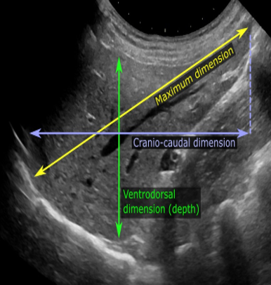
Therefore, a normal liver shows the following sonographic features:
- Homogenous parenchymal echogenicity
- Smooth borders
- Liver normal size less than 17 cm
Liver lobes on ultrasound:
The liver is divided into the right lobe, the left lobe and the caudate lobe. However, knowledge of these lobes is of great importance for the correct ultrasound examination and localization of lesions.
Therefore, it includes the following:
Right lobe on liver ultrasound:
is the largest portion of the liver. however , its includes
- Segments V, VI, VII and VIII
Moreover, we can access the right lobe with an intercostal approach.
Left lobe on liver ultrasound:
is located in the epigastric region. However, it contains
- Segments II, III and IV
Moreover, we can access the left lobe with a subxiphoid approach.
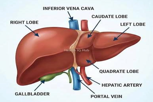
Caudate lobe
In this we can access segment I.
Therefore, found anterior to the IVC and posterior to the portal vein.
Sonographic landmarks
so , identifying sonographic landmarks are essential for accurate diagnosis
Portal Vein in Liver Ultrasound:
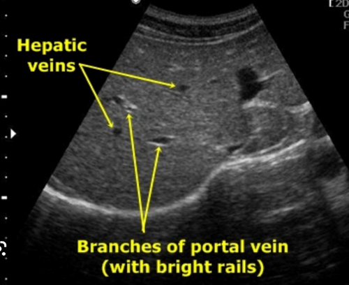
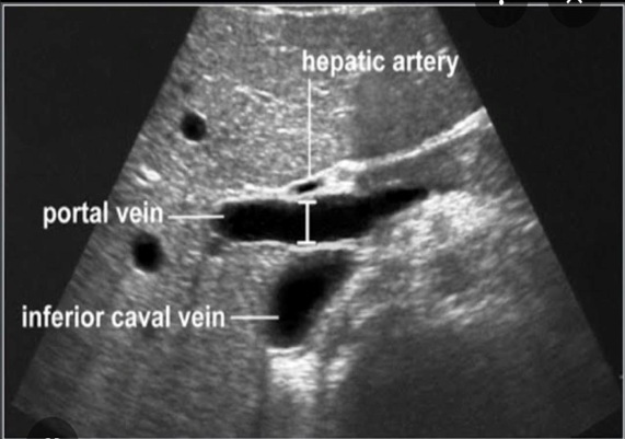
Therefore, among all the vascular structures, the portal vein is the most important landmark
Sono-appearances
- Thick echogenic walls
- Branches into right and left portal veins
Remember: moreover, starry appearances on liver parenchyma
Hepatic veins in liver ultrasound:
However, hepatic veins consist of the following:
- Right hepatic vein
- Middle hepatic vein
- Left hepatic vein
Sono appearances:
Therefore, it appears on ultrasound:
- Thin walls
- Toward IVC
Inferior vena cava:
The portal vein and hepatic veins drain into the IVC.
Sono appearances
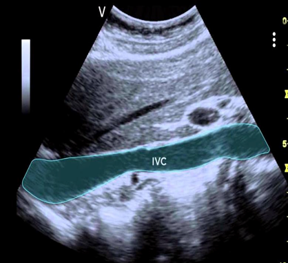
- Thick walls
- Presented vertically and with respiration-change caliber
Ligamentum Teres
however ,it Is remnant of umbilical vein
Sono appearance
- Echogenic band in left lobe
- It’s a landmark for the identifying segment IV
Couinaud segments simplified
Therefore, for anatomical localization liver is divided into eight segments
Segment I
In this caudate lobe is present
Segment -II and III
Left lateral lobe (left of hepatic vein and falciform ligament)
Segment IV
Left medial lobe (between left and middle hepatic veins)
4a- superior (above)
4b-inferior (below)
Segment V and VIII
Right anterior section
V is located below and VIII I located above the portal plane between middle and right hepatic veins
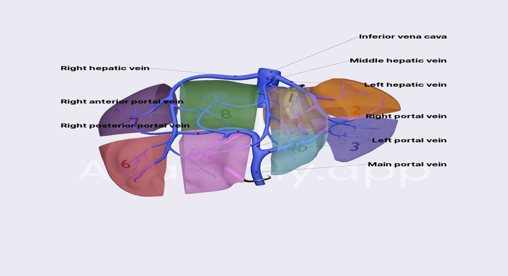
| Memory trick: Clockwise from left lobe II-III-IV-V-VI-VIII |
Segment VI and VII
Right posterior section
Normal Measurements
| Structure | Normal Size |
| Liver length | <15–16 cm |
| Portal vein | <13 mm |
| Common bile duct | <6 mm |
| IVC | Variable |
| Hepatic artery | <7 mm |
Frequently asked questions (FAQs)
Is liver ultrasound used for a variety of reasons?
yes, It is used to assess fatty liver, cirrhosis, liver enlargement, masses and other liver diseases.
Is it necessary to have an ultrasound of the liver before fasting?
Yes, it is usually recommended to fast for 6-8 hours to get better images.
Is liver ultrasound painful?
Not at all, liver ultrasound is safe, painless and non-invasive.
Can liver ultrasound be used to detect fatty liver?
Yes, fatty liver changes are often detected by ultrasound.
On ultrasound, what is the normal size of the liver?
The liver is normally about 15–16 cm long in adults.
Does Doppler play a role in liver ultrasound?
Yes, Doppler plays an important role in evaluating blood flow.


An exceptionally well-written and informative article.. The clear explanations, practical approach, and attention to detail make this one of the most valuable resources on live ultrasound I have come across. Thank you for creating such high-quality educational content.👍
One of the finest educational resources I have read on liver ultrasound. The attention to detail, logical flow, and practical perspective reflect a great deal of expertise and effort. Articles of this quality make complex topics far more approachable and meaningful. Thank you for contributing such valuable knowledge to the medical community.
An exceptional and thoughtfully crafted article on liver ultrasound. The way complex concepts are translated into practical, clinically relevant knowledge is truly impressive. The content is accurate, engaging, and remarkably easy to understand without compromising on depth. Resources like this play an important role in advancing ultrasound education. Thank you for sharing such an outstanding contribution.