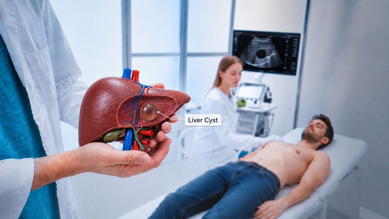
A hepatic cyst ultrasound remains the primary imaging modality for evaluating the characteristics of liver cysts. Because it’s a safe, non-invasive and widely available imaging technique. Additionally, evaluation through sonographic results enables a medical practitioner to differentiate between lesions that need more research.
In order to assist readers, this guide focuses on the imaging characteristics and clinical interpretation of hepatic cysts:
What is a hepatic cyst?
A hepatic cyst is the fluid-filled sac that develops within liver parenchyma. However, liver cysts are benign, non-neoplastic and lined with epithelial cells and filled with clear serous fluid.
Although simple hepatic cysts usually do not cause any symptoms, a correct diagnosis of imaging features is crucial, as some liver cysts can have complex features and need further evaluation. Therefore, careful interpretation plays a crucial role in the final diagnosis.
Why ultrasound is the preferred first-line imaging modality:
Ultrasound is considered a first-line imaging modality; moreover, it provides excellent real-time visualization of hepatic cysts, allowing a detailed analysis of cyst size, walls and internal content. However, if ultrasound shows suspicious features, further evaluation through a CT scan or MRI may be required to make a definitive diagnosis.
As a result, ultrasound clearly demonstrates characteristics. Features of simple hepatic cysts are often easily recognized and can be diagnosed with a high degree of certainty.
Epidemiology of hepatic cyst:
It’s useful in determining the prevalence and distribution of hepatic cysts in the general population. In addition, knowledge of prevalence, demographics and risk factors aids in the interpretation of imaging findings in proper clinical context.
Prevalence of hepatic cysts: Therefore, estimated prevalence is 15-18% in the general population. In addition, their detection has increased due to widespread use of abdominal imaging.
Age factor of hepatic cyst: Although hepatic cysts can occur at any age, they are most frequent in adults over 40 years. Furthermore, clinicians detect these lesions more frequently with increasing age.
Sex factor: However, simple hepatic cysts are more common in females than males. While the exact cause is unknown, researchers suggested hormonal factors.
Risk factors of hepatic cysts: While most simple hepatic cysts are sporadic in nature and have no known cause. Therefore, a number of factors have been linked; some of them are the following:
1- For instance, studies have identified chronic Hepatitis B surface antigen (HBsAg) positivity as an independent risk factor
2-Similarly, individuals with renal cysts are more likely to develop hepatic cysts.
3- Further, metabolic and lifestyle conditions may increase the risk

Clinical presentation of hepatic cysts:
In most cases, hepatic cysts are asymptomatic, and clinicians can discover them incidentally during the abdominal ultrasound. However, a large cyst may produce symptoms by compressing the adjacent organs. Furthermore, complex or complicated cysts can present with additional clinical features.
Therefore, some of them are following:
- Right upper quadrant pain or discomfort
- Occasionally, palpable abdominal mass
- Then, Acute abdominal pain
- Fever and localized tenderness
- Additionally, nausea or abdominal bloating
Sonographic features of hepatic cysts:
While the majority of hepatic cysts are simple and benign, not all hepatic cysts have the same sonographic appearance. Thus, the assessment of the lesion’s echogenicity, wall characteristics, internal architecture and vascularity are critical for correct diagnosis and clinical interpretation. Therefore, clinicians can be confident in differentiating benign from malignant hepatic cysts by recognising the typical features of simple and complex cysts.
How does a simple hepatic cyst appear on ultrasound?
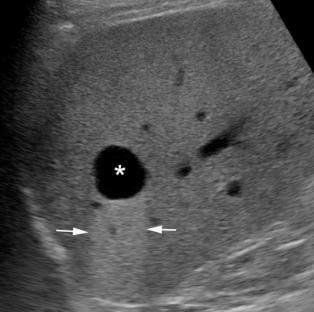
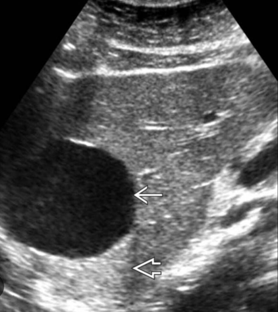
Typically, a simple hepatic cyst is typically sonographically benign and is usually easily diagnosed. Therefore, here are some typical features:
- Anechoic fluid content
- Round or oval shape
- Thin smooth walls
- However, well-defined margins
- No internal echoes
- Additionally, posterior acoustic enhancement
- Therefore, no internal vascularity
Which ultrasound findings suggest a complex hepatic cyst?
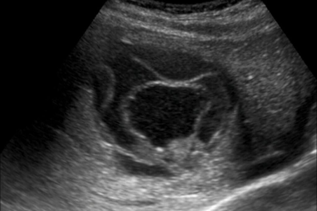
In contrast, a complex hepatic cyst has one or more atypical findings which suggest a haemorrhagic, infectious or cystic neoplasm. Consequently, there is a need to further evaluate these lesions by CT or MRI.
Therefore, here are some atypical features:
- Internal echoes or debris
- Septations
- Thick or irregular walls
- Then, heterogeneous appearance
- Additionally, internal vascularity on color Doppler
Differential diagnosis of hepatic cysts:
While a simple hepatic cyst will have characteristic sonographic features, there are several cystic liver lesions that can have a similar appearance. Thus, a careful analysis of the morphology, echo content and Doppler of the lesion is crucial to an accurate diagnosis.
| Condition | Key USG Features | Differentiating Features |
| Simple hepatic cyst | Anechoic, thin walls, posterior acoustic enhancement | No Septations , no vascularity |
| Hydatid cyst | Daughter cyst, internal membranes | History of exposure or endemic region |
| Liver abscess | Thick walls, internal debris | Fever, sign of infection |
| Hemorrhagic cyst | Internal echoes or clots | Recent pain, intracystic bleeding |
| Biliary cystic neoplasm | Septations or mural nodules | Internal vascularity |
| Cystic metastasis | Irregular cystic lesion | Known primary malignancy |
Ultrasound reporting of hepatic cyst:
A structured ultrasound report provides clear communication between the radiologist and the referring clinician. Therefore, each hepatic cyst must be described systematically, based on its key sonographic characteristics. Furthermore, a description of common benign variants enables differentiation of simple liver cysts from complex cystic liver lesions.
Findings:
A well-defined, round anechoic cyst is seen in the right hepatic lobe (Segment VI) measuring 2.8 × 2.4 × 2.3 cm. In addition, the lesion has a thin, smooth wall and acoustic enhancement in the posterior. Colour Doppler examination fails to show any internal echoes, septations, mural nodules, calcifications, or internal vascularity.
The rest of the liver parenchyma shows normal echotexture and no other focal hepatic lesions. Intrahepatic bile ducts are not dilated, and portal and hepatic veins are not remarkable.
Impression:
- Ultrasound features are suggestive of a typical simple hepatic cyst. There are no suspicious sonographic features
Frequently asked questions (FAQS)
Can a hepatic cyst rupture?
A hepatic cyst can rupture and cause sudden abdominal pain.
Can a hepatic cyst cause abdominal pain?
Yes, large hepatic cysts may cause a right upper abdominal pain and discomfort.
When should a CT or MRI follow a hepatic cyst ultrasound?
CT or MRI should follow when ultrasound shows a complex cyst.
Can a hepatic cyst grow over time?
Yes, some hepatic cysts grow gradually in size.


Excellent breakdown of hepatic cyst ultrasound findings. I especially liked how the article explained key sonographic features in a clear and practical way. Highly recommended…✔️💯
Excellent
Very well explained 😃 keep it up,✅