
Spleen ultrasound is one of the most widely used diagnostic imaging modalities for evaluating the spleen due to its safety, accessibility and ability to provide real-time visualisation without ionising radiation. Additionally, it is a cost-effective and non-invasive imaging technique. In addition, this comprehensive guide covers the indications, patient preparations, sonographic anatomy, scanning protocol and normal measurements of spleen ultrasound in clinical practice
What is a spleen ultrasound?
Spleen ultrasound (splenic ultrasonography) is a diagnostic ultrasound of the spleen. Furthermore, the scanning can help measure the size, shape, and echogenicity along with blood flow of the spleen.
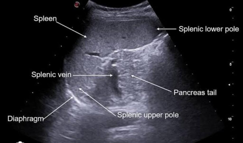
Normal spleen ultrasound appearance: spleen appear homogenous
Clinical indications for spleen ultrasound:
Spleen ultrasound is done to look at the spleen and find problems. In addition, it may identify the size, structure, blood flow or how well the spleen is working:
When is spleen ultrasound recommended?
- Detect splenomegaly (enlarged spleen) and determine its etiology
- Similarly, evaluate the splenic injury in blunt abdominal trauma / accidents
- Additionally, diagnose splenic cysts, abscesses and focal masses
- Furthermore, Doppler ultrasound for diagnosis of splenic infarction and blood flow abnormalities
- Moreover, access for left upper quadrant abdominal pain or tenderness with splenic disease
- likewise , Evaluate for splenic involvement in hematological disorders (anaemia, leukaemia, lymphoma)
- Examine the spleen for enlargement in the context of liver cirrhosis and portal hypertension
- furthermore , check abnormal blood cell counts if you suspect a problem with the spleen
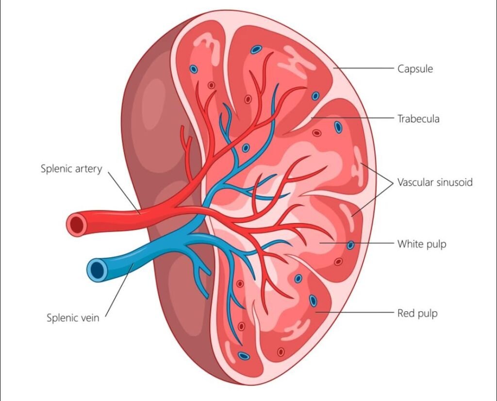
Patient preparations for spleen ultrasound:
An accurate spleen ultrasound examination is ensured by proper patient preparation, which also helps to increase image quality. Although fasting is not required for splenic ultrasound, the patient should:
- Drink water as advice if further abdominal organs are being evaluated
- Next, for simple access to upper abdomen dress comfortably and loosely
- Finally, inform the sonographer about any recent abdominal surgery, trauma, or our previous medical history
Spleen Ultrasound Scanning Techniques:
A systemic evaluation of the spleen is essential for proper visualisation of the spleen. Moreover, following appropriate protocols may improve diagnostic accuracy.
Position patient for splenic ultrasound:
- Supine and right lateral decubitus positions are used for scanning
- However, if visualisation is limited Ask the patient to take a deep inspiration for better visualization of spleen beneath the rib cage
Probe Selection for Splenic Ultrasound:
- Make sure us of curvilinear transducer operating at 2-5 MHz
- However a higher frequency linear probe may be utilized for superficial examination
Probe Placement:
- Through the lower intercostal gaps, place the transducer across the left upper quadrant
- Next, obtain longitudinal and transverse views of the spleen
- Finally evaluate the splenic hilum, surrounding organs and adjacent vascular structures
Common Technical Challenges:
Several factors may limit the splenic visualisation.
- higher BMI (Obesity)
- Overlying bowel gass
- Rib shadowing
- Shallow breathing
Image Optimization Tips:
- Use intercostal approach to reduce rib shadowing
- Furthermore, adjust depth so the spleen occupies most of screen
- Moreover, optimise overall gain and time gain compensation
- Additionally, guide the patient to hold a deep inspiration for better visulization
Sonographic anatomy:
Knowing the sonographic anatomy of the spleen is crucial for correct interpretation of ultrasound examinations. In addition, knowledge of surrounding anatomical landmarks assists sonographers to identify the spleen and detect pathological changes with more confidence.
spleen on ultrasound: here is complete guide about the spleen on ultrasound, including location, shape, size, etc.
Location of Spleen:
- Left upper quadrant
- Beneath the left hemidiaphragm
- Posterior to stomach
- Adjacent to ribs 9-11
Shape and Surface:
The normal spleen has a crescent-shaped or wedge-shaped appearance with a smooth outer contour. It consist of
- Convex diaphragmatic surface
- Concave visceral surface
- Superior border
- Inferior border
- Medial splenic hilum
(The splenic hilum serve as the entry and exist point for splenic artery, vein and nerve.)

Echogenicity and Echotexture:
A healthy spleen appears with a uniform and homogenous echotexture. However, normally spleen parenchyma shows the following:
- Slightly hyperechoic then liver
- More echogenic then renal cortex
- Furthermore, homogeneous without focal lesions
- Moreover, smooth without capsular irregularity
Splenic Hilum:
The splenic hilum is located on the medial surface of the spleen and represents the important sonographic landmark. It contains
- Splenic artery
- Splenic vein
- Lymph vessels
- Autonomic nerves
During ultrasound examination, identifying the splenic hilum will help to diagnose vascular abnormalities.
Splenic Vascular Anatomy:
The spleen has richest blood supplies in the body
The splenic artery, a branch of celiac truck, coursed along the superior border of the pancreas before entering the splenic hilum.
The splenic vein exits the hilum and joins the superior mesenteric vein from the portal vein.
Furthermore, Doppler ultrasound allows assessment of:
- Splenic artery patency
- Splenic vein patency
- Portal venous circulation
- Splenic perfusion
Adjacent Anatomical Landmarks:
- Left kidney
- Left hemidiaphragm
- Stomach pancreatic tail
- Splenic flexure of colon
Normal Sonographic Appearance:
- Smooth margins
- Homogenous echotexture
- Mildly hyperechoic to liver
- Moreover, no focal masses
- Normal vascularity
Normal Measurements
| Structure | Measurement |
| Length | 12 cm |
| Width | 6-8 cm |
| Thickness | 3-5 cm |
| Splenic vein approx. | 8-10 mm |
Spleen ultrasound is the safe, noninvasive and cost-effective imaging modality; however, it is used to evaluate the size, structure, echogenicity and vascularity. Additionally, it plays an important role in diagnosing splenomegaly, traumatic injuries, and focal splenic lesions. Overall spleen ultrasound remains a non-invasive imaging modality for clinical practice.
Frequently asked questions (FAQs)
1- Does fasting require it before a spleen ultrasound?
Fasting is usually not required for an isolated spleen but may be recommended due to scanning of other abdominal organs.
2-How long does a spleen ultrasound take?
It usually takes 15-30 minutes for a complete examination.
3- Is splenic ultrasound invasive?
No, it’s non-invasive and safe for diagnosis.
4-Why would a doctor recommend a splenic ultrasound?
A doctor may recommend the ultrasound to investigate the abdominal pain, splenomegaly, infection or trauma.
5. Which is better for evaluating the spleen: ultrasound or CT scan?
Ultrasound is performed first as an imaging test to evaluate the splenic conditions, while a CT scan provides the evaluation of complex cases after trauma.


One of the most comprehensive and easy-to-follow articles I have read on spleen ultrasound. The practical insights and clear presentation are highly appreciated.
This article on spleen ultrasound is exceptionally well presented. I really liked how the information is explained in a simple yet detailed way, making it easy to understand and apply. The practical approach and clear organization make it a valuable learning resource for anyone interested in ultrasound. ✔️