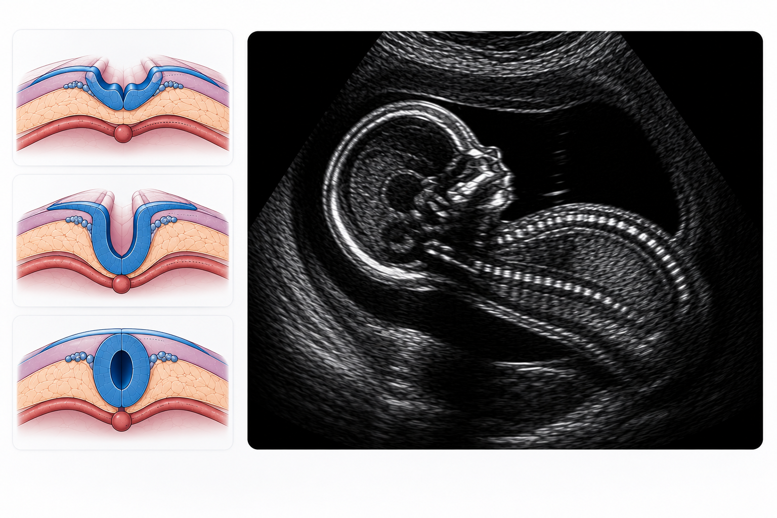
Neural tube defects are one of the most significant congenital anomalies assessed by prenatal ultrasound. However, in my obstetrical ultrasound training, I was taught that even if you see something that is abnormal but small, it can alter the entire approach to a clinical situation. In obstetrics training, I was taught that even if you see something that is abnormal but small, it can alter the entire approach to the clinical situation.
So, experience taught an important lesson: that a successful prenatal ultrasound is not just about the detection of abnormalities but also their clinical significance. Moreover, this guide explains the structured ultrasound approach.
What are neural tube defects?
Neural tube defects are serious birth defects that occur when the neural tube does not close completely during the third and fourth weeks (22-28 days) of conception and pregnancy. Therefore, the brain, spinal cord and vertebrae developed abnormally. However, the neural tube closes early in pregnancy, so many affected pregnancies are already in progress before the mother realize that she is pregnant.
As I was doing my fetal ultrasound rotations early on, one of my instructors kept saying that it was very important to know what is normal so you could see what is abnormal. Therefore, I came to the realization over the years that this concept is true for nearly all fetal abnormalities during USG exams.
Why early prenatal ultrasound matters:
Prenatal ultrasound is the first-line imaging modality for the detection of neural tube defects. Furthermore, early diagnosis facilitates timely referral and appropriate management of pregnancy and subsequent imaging that is required.
While training to become a sonographer, I was taught never to access the foetal spine without assessing the rest of the body. Therefore I always evaluate skull, brain, posterior fossa and spine as a unit
Benefits of prenatal ultrasound:
Moreover, the benefits of early ultrasound are numerous. Therefore, here are some benefits of prenatal ultrasound:
- However, identify large neural tube defects before birth
- Then, systematically access the fetal brain and spine
- Improve prenatal counseling
- Provide multidisciplinary pregnancy management
- Therefore, provide direction for fetal MRI or genetic testing
Normal fetal neural tube anatomy:
However, all sonographers need to know the normal development of the foetal neural tube. Therefore, routine obstetrics makes it easier to identify fetal abnormalties.
Embryology of neural tube:
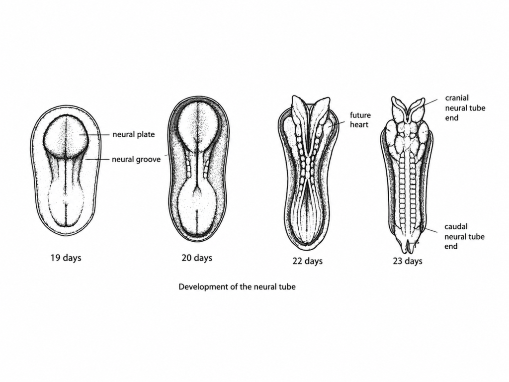
Although the embryonic ectoderm forms the neural tube soon after fertilization. The neural folds then fuse together to form the neural tube
Therefore this structure eventually becomes the
- Brain
- Brain stem
- Spinal cord
Normal neural tube closure:
The normal tube closure takes place between day 22 and day 28 post fertilization. Moreover, at this time both the cranial and caudal neuropores are completely closed.
However, if closure remains incomplete, neural tissue becomes exposed. As a result, birth defects like spina bifida or anencephaly can develop.
Fortunately, sufficient folate intake by the mother before becoming pregnant and during early pregnancy can greatly lower the risk of neural defects.
Systemic ultrasound assessment:
However, fetal anomaly scan is a crucial time to identify neural defects and requires a systemic ultrasound evaluation. Therefore, sonographers should not look at the spine but also at the fetal central nervous system in systemic order.
Moreover, this method helps to minimize the likelihood of missing abnormalities and increase the confidence of diagnosis.
Evaluate the Skull
Access the foetal skull first in axial and then in sagittal. Examine the cranial vault for integrity, shape and contour. Therefore, search for calvarial defects or the lemon sign.
Evaluate the brain.
Then explore the foetal brain with the typical sections. Assess the cerebral hemispheres, lateral ventricles, and cavum septi pellucidi and midline structure. In addition, examine the ventricles, as they can be associated with open neural tube defects.
Evaluate the Posterior Fossa
After that, evaluate the posterior fossa thoroughly. Observe the shape of the cerebellum and measure the cerebellum and cisterna magna. Also check for the banana sign, which is a well-known indirect sign of open spina bifida.
Evaluate the spine
Next look for the foetal spine in the sagittal, transverse and coronal planes. Evaluate the relationship between the alignment of the vertebral bodies, posterior ossification centres and overlying skin. In addition, look for any discontinuity, widening, cystic protrusion or exposed neural tissue.
Search for associated anomalies
Lastly, conduct a thorough examination for any other foetal defects. Neural tube defects often occur in association with other congenital defects, so assess the ventricles, kidneys, heart, limbs and amniotic fluid volume. Thus, the ability to recognize associated findings will enhance the accuracy of diagnosis and help to guide prenatal counselling.
Sonographic features of neural tube defects:
The sonographic findings can be divided into direct ultrasound findings, where the defect itself is seen, and indirect cranial signs which are caused
Direct ultrasound findings
Direct ultrasound findings show the main abnormality of the fetal skull or spine. These results usually help to confirm the diagnosis of a neural tube defect and its site.
Cranial defects: evaluate the fetal calvarium, brain tissue and cranial contour such as
1-Acrania: Absence of skull bone (brain matter moves freely because there is no vertex on the skull). And giving the Mikey mouse sign (bulging cerebral hemispheres)
2-Anencephaly: The first detected abnormality where a baby is without parts of the brain and an absent skull bone. The frog eye sign / owl eye sign seen on ultrasound
3-Microcephaly: In this condition, head circumference decreases by three standard deviations.
4-Dolicocephaly: an oval-shaped head increase in length while decreasing in width
5-Branchiocephaly: In this condition the width of the head increases while the length decreases.
6-Microcephaly: The condition in which head circumference increased by two standard deviation
Spinal defects: look for defects in posterior vertebral elements, widening of the spinal canal, discontinuity of skin, cystic protrusion or exposure to neural tissue.
1-Meningocele: Cystic sac protrude through vertebral defect (intact overlying skin in most cases) and normal spinal cord remain within the spinal canal
2-Myelomeningocele: protrusion of the meninges and spinal cord defects through a vertebral defect. This portion of spinal column allow the spinal cord to protrude through the opening
- Associated with Alnorld Chiri Malformation:
Indirect Cranial Sign
Lemon-shaped head: bilateral scalloping of frontal bones
Banana shape: cerebellum (normally, the cerebellum is dumbbell-shaped)
Venticulomegaly: lateral ventricles diameter more than 10 mm
Ultrasound Features by Pathology:
Spina Bifida: Any closing or formed defect in the spine lead to spina bifida.
Occulta
Anencephaly:
- Absent calvarium and most of brain
- Rudimentary brain tissue visible
- Bulging fetal orbits giving the characteristic owl-eye or frog-eye appearance
- Increase fetal activity because of irritating effects of amniotic fluid on exposed meninges and neural tissue
- Hydramnios occurs in 50% after 25 weeks of gestation
- Coexisting spina bifida is also common with anencephaly
Encephalocele:
- The ultrasonography appearance of a posterior Encephalocele defect characterized by sac-like protrusions of the brain and membrane that cover it through opening in the skull
- Commonly involved with the occipital bone are usually located in the midline.
Differential Diagnosis
| Acrania | Absent of skull bone |
| Anencephaly | Absent of skull bone and brain matter |
| Encephalocele | Skull defect with herniation of brain tissue and/or meninges |
| Ventrecolomegaly | CSF increase in ventricle |
| Spina Bifida | Any closing or forming defect in spine |
| Meningocele | Cystic sac containing only meninges through spinal defect |
| Myelomeningocele | Cystic sac containing meninges and vertebrae through spinal defect |
| Holoprosencephaly | If there is any defect in the flax line, proboscis, or cyclopia. Arhinia, Cleft lip, single nostril |
Ultrasound Reporting Checklist
Include
- Gestational age
- Lesion type, size, skin covering /not
- Cranial findings
- Ventricular size is normally less than 10 mm.
- Posterior fossa
- Associated anomalies
- Amniotic fluid In the Phelan method, the normal AFI is between 9 and 19
- Placenta: The size of the placenta correlates with the gestational age till 28 weeks. If the size of placenta exceed 45 mm, it leads to placentomegaly
Recommended follow-up (foetal MRI, genetic counseling)
Frequently asked questions (FAQs)
1-Is ultrasound able to detect neural tube defects in an unborn baby?
Yes, most neural tube defects can be detected in the first or second trimester with prenatal ultrasound.
2-What is the time of diagnosis for neural tubes on ultrasound?
Most neural tube defects are detected during the 18-22 week fetal anomaly scan.
3. Which of the following is the most common neural tube defect found on ultrasound?
The most common neural tube defect detected by prenatal ultrasound is spina bifida.
4-is folic known to prevent neural tube defects?
Yes, folic acid before pregnancy and early pregnancy is very effective at decreasing the risk of neural tube defects