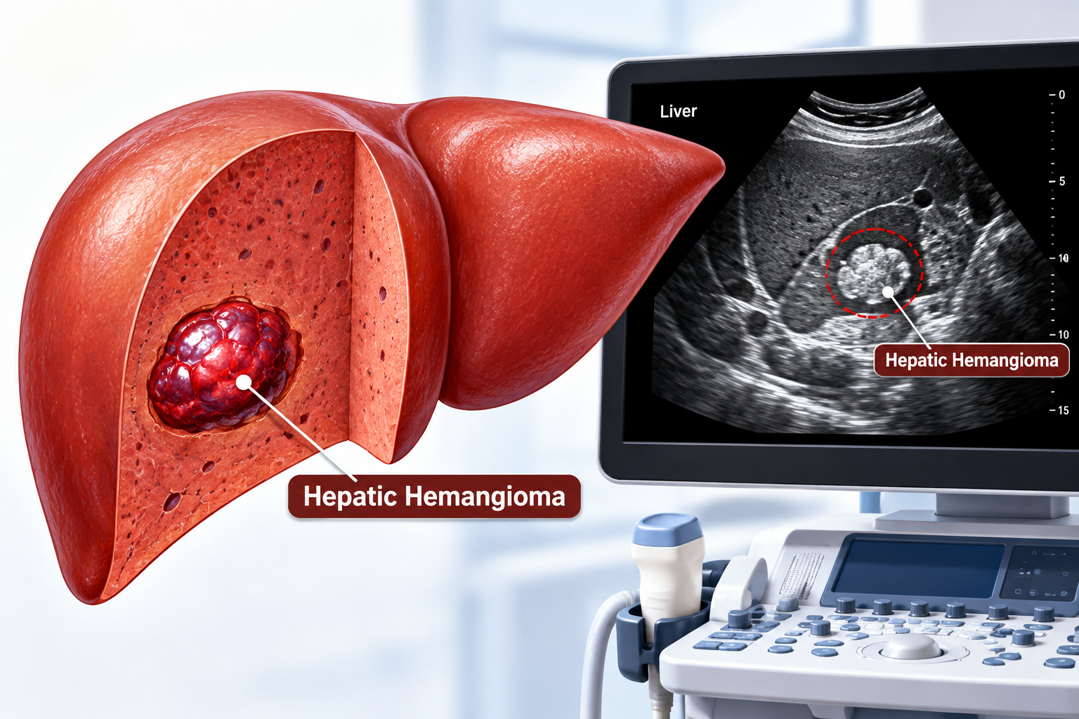
Liver hemangioma ultrasound is an important part of sonographic evaluation of focal liver lesions. However, hepatic haemangiomas usually appear well defined and homogenous, with a characteristic of hyperechoic appearance. Atypical appearance can make diagnosis difficult.
Hence, the evaluation of echogenicity, margins, size, Doppler vascularity and background liver are essential for proper interpretation and reporting
What is liver hemangioma?
The most common benign vascular liver lesions are hepatic hemangiomas or hepatic malformations. Moreover, patients have not usually associated with any symptoms. Therefore, it’s important to differentiate hemangiomas from other hepatic malignancies in a sonography setting.
Prevalence: 0.4% – 20% of the general population.
Number: most are solitary.
Location: usually found in the right lobe of the liver
Clinical features of hemangioma:
- Most liver hemagiomas cause no symptoms, and doctors detect them incidentally during ultrasound or other imaging
- Typically they usually appear at any age but are more common in adults (30-50) and more common in females (M:F 5:1).
- Additionally, hormonal factors can affect the behaviour of lesions: some patients have had an increase in the size of hemangiomas with estrogen exposure (during pregnancy/estrogen therapy)
If symptoms appear may include:
- Right upper quadrant pain
- Reduced appetite
- Early satiety
- Abdominal fullness
- Nausea or vomiting
Case spotlight:
A 26-year-old female comes to the radiology department with complaints of upper abdominal pain associated with nausea and vomiting. Clinicians requested abdominal ultrasound to investigate her symptoms.
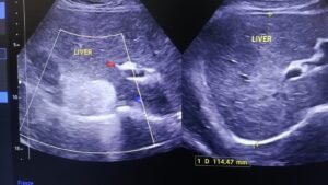
Ultrasound findings:
The liver is normal in size, with mildly inversed echogenicity of liver parenchyma suggestive of hepatic steatosis.
A well-defined, homogenous, smooth-margined and predominantly hyperechoic focal liver lesion was noted within the liver. Mild posterior acoustic enhancement was noted. No colour flow was seen on CDI.
Opinion:
- Ultrasound features are suggestive of hepatic hemangioma
What does liver hemangioma look on ultrasound?
Ultrasound is frequently the first imaging modality to be used to assess focal liver lesions; however, its availability, non-invasiveness and ability to assess the liver parenchyma in real time.
On ultrasound, a typical liver hemangioma will show the following characteristics:
Echogenicity: homogenous and hyperechoic to the adjacent liver
Margins: well defined, helps to differentiate it from poorly defined focal lesions
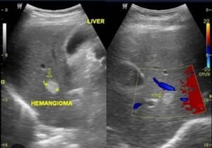
Shape: round or oval, most of time
Echotexture: usually uniform, especially in small lesions; however, larger hemangiomas may develop into heterogeneous tumours.
Posterior features: however, posterior acoustic enhancement may be present.
Size: therefore, the majority of hemangiomas are small, usually less than 3 cm.
But does every liver hemangioma have this classic ultrasound appearance? Not necessarily. Although most typical lesions demonstrate characteristic sonographic features, larger or complicated hemangiomas may appear differently. Therefore, distinguishing typical from atypical findings is essential for accurate interpretation.
Liver hemangioma on colour Doppler:
- However, Hepatic hemangioma typically show no internal blood flow on colour Doppler ultrasound because blood flow inside the hemangioma is extremely slow.
- Sometimes colour Doppler may show the small feeding blood vessels around the other edge (peripheral blood flow)
What does hepatic hemangioma look like on ultrasound when patients have fatty liver?
Hepatic steatosis has become more common in recent years. However, hepatic steatosis makes the background of the liver brighter than usual. Therefore, in these cases, hepatic hemangioma seem less echogenic and hypoechoic than the surrounding liver.
Frequently asked questions:
1-is it possible for liver hemangioma to be hypoechoic?
Yes. There are atypical appearances, especially related to the characteristics of the lesions and background liver.
2- Is there blood flow within a liver hemangioma on Doppler?
Usually little or no detectable internal flow is seen on conventional Doppler.
3- On ultrasound, what does a liver hemangioma appear as?
Usually a well-defined, homogeneous hyperechoic lesion
4-Is it possible to diagnose liver hemangioma by ultrasound?
A typical lesion in a suitable clinical context can be confidently diagnosed on ultrasound, and if the lesion is atypical, further imaging may be necessary.