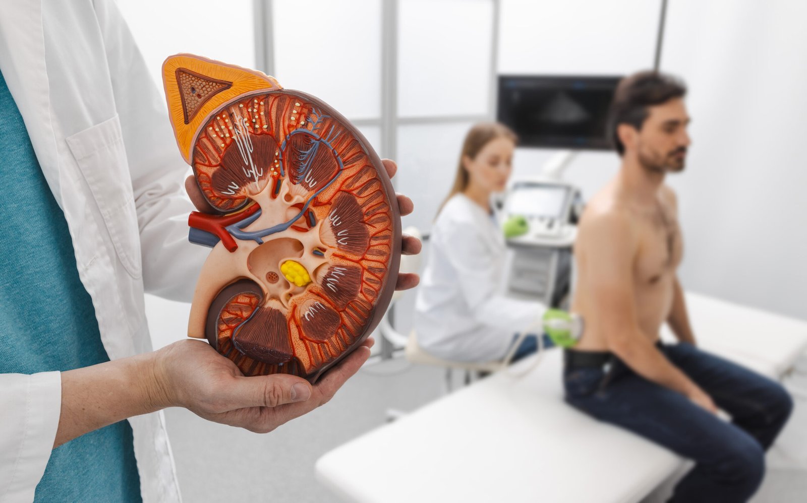
(Learn how to do kidney ultrasounds and preparations to interpretation)
Ultrasound is a diagnostic imaging method that uses sound waves to create the image of organs inside the body. It produces real-time images without radiation; it provides fast, accessible and cost-effective clinical assessment. It’s usually used to evaluate the abdominal, pelvic, renal and vascular conditions.
What is a kidney ultrasound?
Kidney ultrasound (renal ultrasound 1) is an imaging technique that is used to evaluate renal anatomy and pathology. It helps healthcare professionals to access the kidney size, structure and overall conditions without exposing them to radiations.
Why is kidney ultrasound performed?
Kidney ultrasound is performed to evaluate the kidney and identify the conditions that may affect the kidney function or overall health
- Kidney ultrasound may be recommended to:
- Detect kidney stones (renal calculi) or urinary tract obstruction
- Evaluate hydronephrosis (kidney swelling) caused by impaired urine drainage
- Identify renal cysts, masses and tumors
- Evaluate the cause of blood in urine (hematuria)
- Examine possible cause of the flank pain and recurrent urinary tract infection
What Patient preparations for kidney ultrasound:
- Maintain adequate hydration before examination unless instructed
- The patient may be advised to drink water and retain urine to ensure the optimal bladder distension when urinary tract assessment is required
- Wear lose and comfortable cloths to facilitate to abdomen during kidney ultrasound
- Before performing a renal ultrasound scan, all available clinical histories, imaging examination results, and laboratory test results must be provided.
Scanning techniques for kidney ultrasound:
Kidney ultrasound (renal sonography) is performed with a curvilinear low-frequency probe (2-5 MHz) to obtain optimal penetration and resolution of deep abdominal structures.
Patient positioning:
- Patient is typically scanned in supine position as a baseline approach
- During the scan, the patient is positioned in the lateral decubitus and oblique positions
- inspiration may be requested to displace the bowel gases and improve acoustic window
Scanning planes and techniques:
The examination includes the assessment of both kidneys in longitudinal and transverse planes with careful adjustment of probe position and patient orientation for optimised image quality. Real-time adjustment of gain, depth and focal zone is performed to obtain the image quality.
Right kidney
- Orient the probe marker toward the patient’s head
- For longitudinal view Place the probe at right mid-axillary line, typically between the 10th and 11th intercostal spaces.
- For transverse view adjust the probe orientation counter 90 degree anticlockwise
- Ultrasound probe is moved systematically from the upper renal pole to the lower renal pole to
- Centre the kidney on the ultrasound screen
- Perform slow fanning (tilting anteriorly and posteriorly) to evaluate the entire renal contour and renal parenchyma
Left kidney
- For longitudinal view place the probe in the left flank region approximately 8-10th intercostal space
- For transverse view rotate the probe 90 degree anticlockwise.
- A posterior approach may be required
- And deep inspiration may be is performed due to splenic shadowing
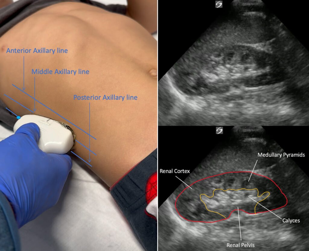
Sonographic anatomy of kidney:
The sonographic anatomy of the kidney is essential for proper interpretation of renal ultrasound. The kidneys are bean-shaped retroperitoneal organs located on either side of the spine, typically between the T12-L3 levels, with the right kidney positioned slightly lower due to the liver.
On ultrasound imaging, the kidneys is evaluated in terms of shape, size, echogenicity and internal
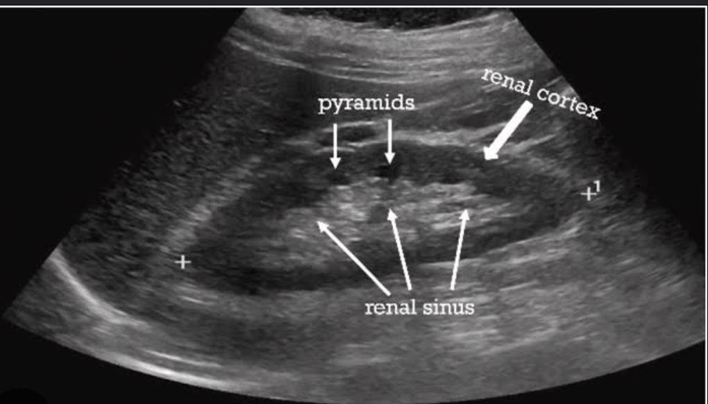
Renal cortex: the outer part of the kidneys, which is homogenous, low to intermediate echoic and slightly hypoechoic to the liver or spleen in a normal adult.
Renal medulla (pyramids): Hypoechoic triangular structures that extend to the renal sinus. These are normally less echogenic than the cortex.
Corticomedullary differentiation (CMD): important to distinguish cortex from medulla, which is a key sign of normal renal parenchyma.
Renal sinus: central echogenic area made up of fat, lymphatic tissue and collecting system structures. It is very echoreflective on ultrasound.
Renal pelvis and calyces: not normally prominent and collapsed unless dilated. In the presence of hydronephrosis, they are seen.
Adjacent structures (acoustic windows):
Right kidney: liver acts as acoustic window for optimal visualisation.
Left kidney: spleen serve as the primary window
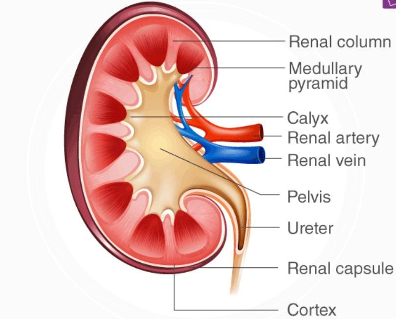
Image evaluation protocols:
- Measurement of renal length in longitudinal plane (upper to lower pole)
- Evaluation of the liver and spleen echogenicity
- Differentiation of Corticomedullary differentiation (CMD)
- Renal sinus, renal pelvis, and calyces are assessed for any dilation
Normal Measurements:
| Parameter | Normal Measurement |
| Kidney Length | 9–12 cm |
| Kidney Width | 4–6 cm |
| AP Thickness | 3–5 cm |
| Cortical Thickness | 6–10 mm |
Frequently asked question:
- Is kidney ultrasound painful?
No kidney ultrasound is a painless and noninvasive procedure.
- How long does kidney ultrasound take?
A routine kidney ultrasound typically take 10 to 15 minutes
- Can I eat before kidney ultrasound?
Yes, kidney ultrasounds don’t require fasting until combined abdominal imaging
- Is there radiation in ultrasound?
No, ultrasound use the sound waves and does not involve the ionizing radiations, making it safe imaging modality
- What happened after the kidney ultrasound?
The image are reviewed and interpreted by radiologists, who provide the findings to healthcare professionals